著名医師による解説が無料で読めます
すると翻訳の精度が向上します
Atrophie Blanche(AB)は、Livedoid血管障害として知られる臨床病理学的存在に続発する瘢痕の形態学的パターンです。それは、下肢、足首、または背側の再発性、痛みを伴う、潰瘍として存在する慢性状態であり、その後の瘢痕形成を伴う若年から中年の女性で主に発生します。Atrophie Blancheは、治癒した潰瘍の結果を説明しています。これらは、末梢の毛細血管ectasiasを伴う白い萎縮性の星瘢痕として特徴的に存在します。Atrophie Blancheは、最も純粋な形では、主要な潰瘍性病変であるLivedoid血管障害としても知られています。生まれた血管障害はあまり理解されていません。それは、その後の皮膚潰瘍を伴う表在性真皮の血管の閉塞を伴うと考えられています。分節性ヒアリン化血管炎、夏の潰瘍を伴うLivedo reticularis、下肢(紫)の網状パターンを伴う痛みを伴う紫斑潰瘍など、この状態を説明する他のあまり一般的に使用されていない用語がいくつかあります。身体検査の所見は疾患の段階に依存しますが、パンチアウトされた潰瘍を発症する両側で痛みを伴う紫斑病の病変を持つ中年の女性には古典的に存在します。これらの潰瘍は慢性で、癒しが遅く、古典的な星の白い瘢痕を残します。それは中年の女性でより頻繁に発生しますが、男性で発生し、小児には病気のいくつかの症例報告があります。他の多くの病気は、アトロフィー・ブランシュと同様のエンドステージを呈するかもしれません。Atrophie Blancheのような病変の原因には、皮膚小血管血管炎と抗リン脂質抗体症候群が含まれます。これらの他の障害は、歴史に先行するパンチアウトされた潰瘍が欠けているため、Atrophie Blancheと区別できます。
Atrophie Blanche(AB)は、Livedoid血管障害として知られる臨床病理学的存在に続発する瘢痕の形態学的パターンです。それは、下肢、足首、または背側の再発性、痛みを伴う、潰瘍として存在する慢性状態であり、その後の瘢痕形成を伴う若年から中年の女性で主に発生します。Atrophie Blancheは、治癒した潰瘍の結果を説明しています。これらは、末梢の毛細血管ectasiasを伴う白い萎縮性の星瘢痕として特徴的に存在します。Atrophie Blancheは、最も純粋な形では、主要な潰瘍性病変であるLivedoid血管障害としても知られています。生まれた血管障害はあまり理解されていません。それは、その後の皮膚潰瘍を伴う表在性真皮の血管の閉塞を伴うと考えられています。分節性ヒアリン化血管炎、夏の潰瘍を伴うLivedo reticularis、下肢(紫)の網状パターンを伴う痛みを伴う紫斑潰瘍など、この状態を説明する他のあまり一般的に使用されていない用語がいくつかあります。身体検査の所見は疾患の段階に依存しますが、パンチアウトされた潰瘍を発症する両側で痛みを伴う紫斑病の病変を持つ中年の女性には古典的に存在します。これらの潰瘍は慢性で、癒しが遅く、古典的な星の白い瘢痕を残します。それは中年の女性でより頻繁に発生しますが、男性で発生し、小児には病気のいくつかの症例報告があります。他の多くの病気は、アトロフィー・ブランシュと同様のエンドステージを呈するかもしれません。Atrophie Blancheのような病変の原因には、皮膚小血管血管炎と抗リン脂質抗体症候群が含まれます。これらの他の障害は、歴史に先行するパンチアウトされた潰瘍が欠けているため、Atrophie Blancheと区別できます。
Atrophie blanche (AB) is a morphologic pattern of scarring that occurs secondary to a clinicopathologic entity known as livedoid vasculopathy. It is a chronic condition that presents as recurrent, painful, ulcers of the lower leg, ankle, or dorsal foot, predominately occurring in young to middle-aged females with subsequent scar formation. Atrophie blanche describes the result of healed ulcers. These characteristically present as a white, atrophic stellate scar with peripheral telangiectasias. Atrophie blanche is also known as livedoid vasculopathy, which in its purest form, is the primary, ulcerated lesion. Livedoid vasculopathy is poorly understood. It is believed to involve occlusion of the blood vessels in the superficial dermis with subsequent skin ulceration. There are several other less commonly used terms to describe this condition, such as segmental hyalinizing vasculitis, livedo reticularis with summer ulceration, and painful purpuric ulcers with reticular patterning on the lower extremities (PURPLE). Physical exam findings will depend on the disease stage but classically present in a middle-aged woman with bilateral, painful, purpuric lower leg lesions that develop punched-out ulcerations. These ulcers are chronic, slow to heal, and leave the classic stellate, white scars. Although it occurs more frequently in middle age females, it does occur in males, and there are a few case reports of disease in children. Many other diseases may present with a similar end-stage as atrophie blanche. The causes of atrophie blanche-like lesions include cutaneous small-vessel vasculitis and antiphospholipid antibody syndrome. These other disorders can be distinguished from atrophie blanche as the history will lack preceding punched-out ulceration.
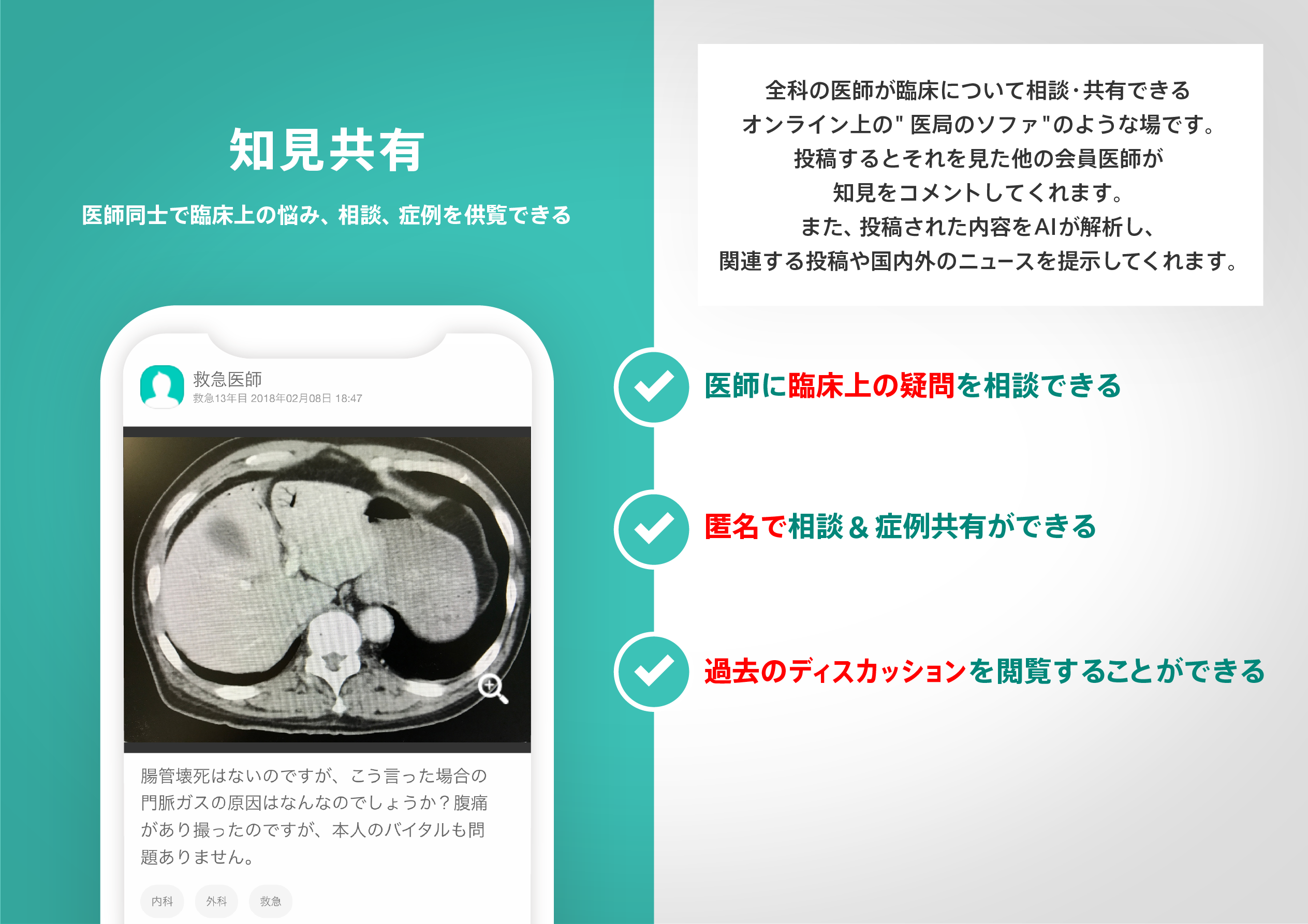
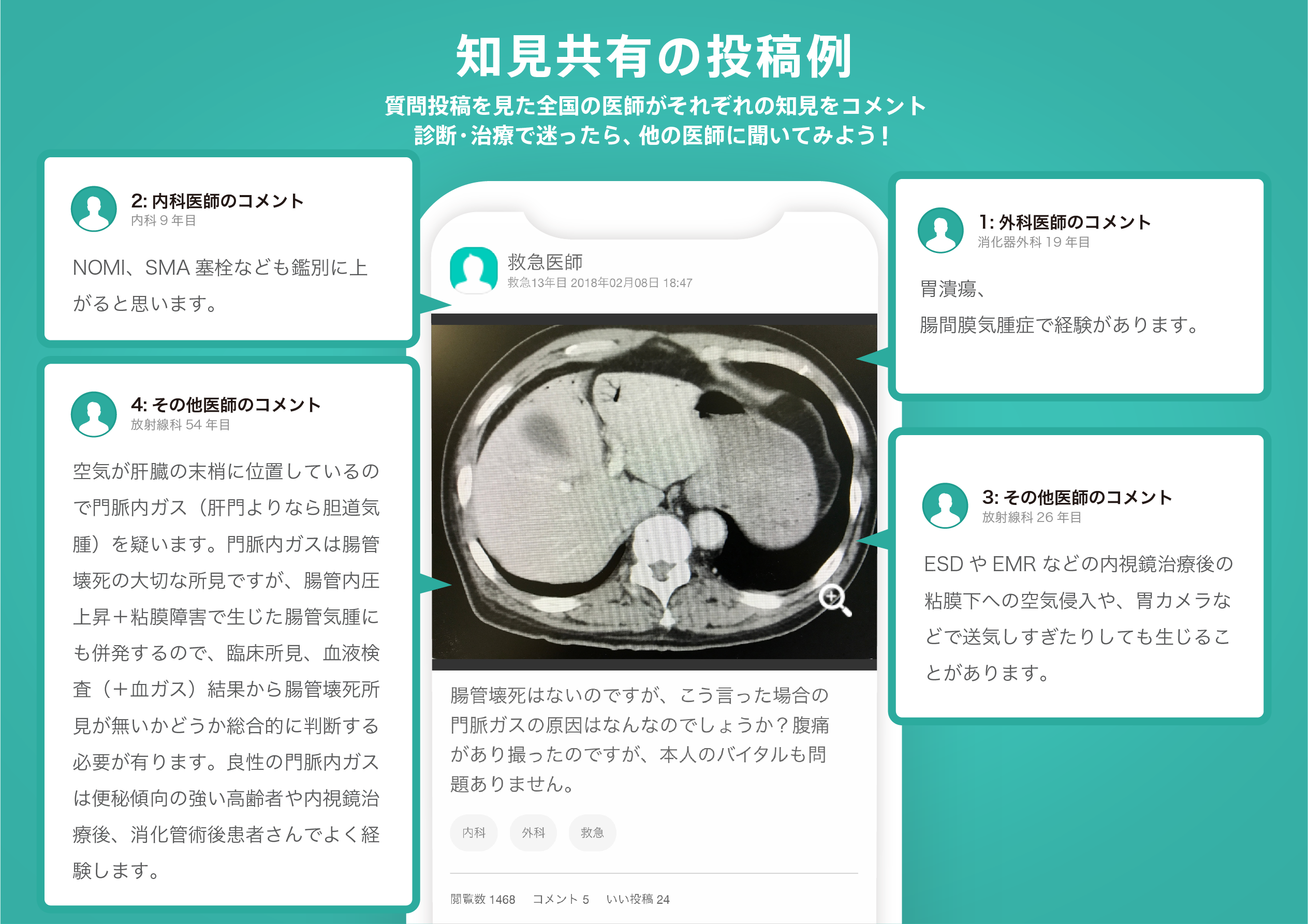
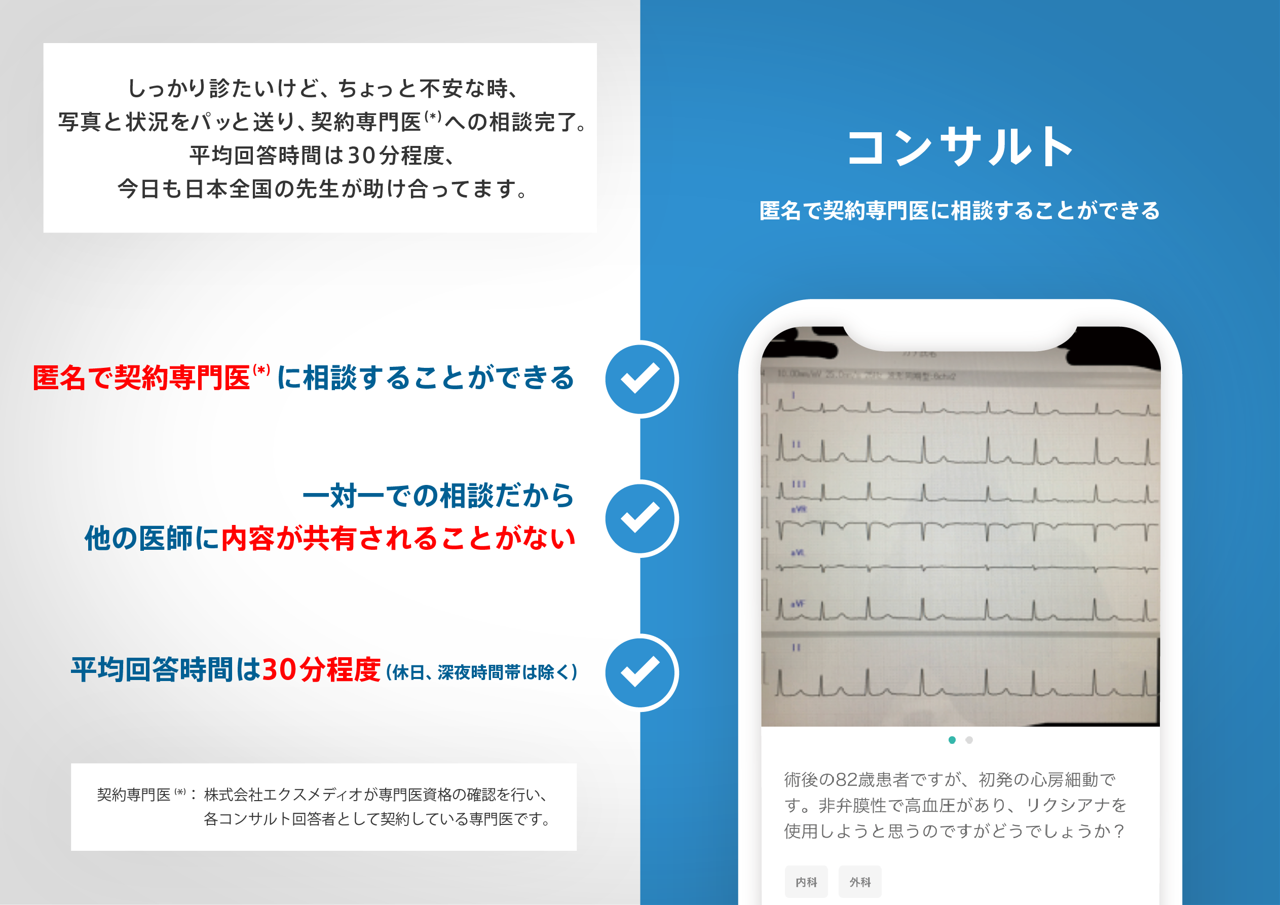
医師のための臨床サポートサービス
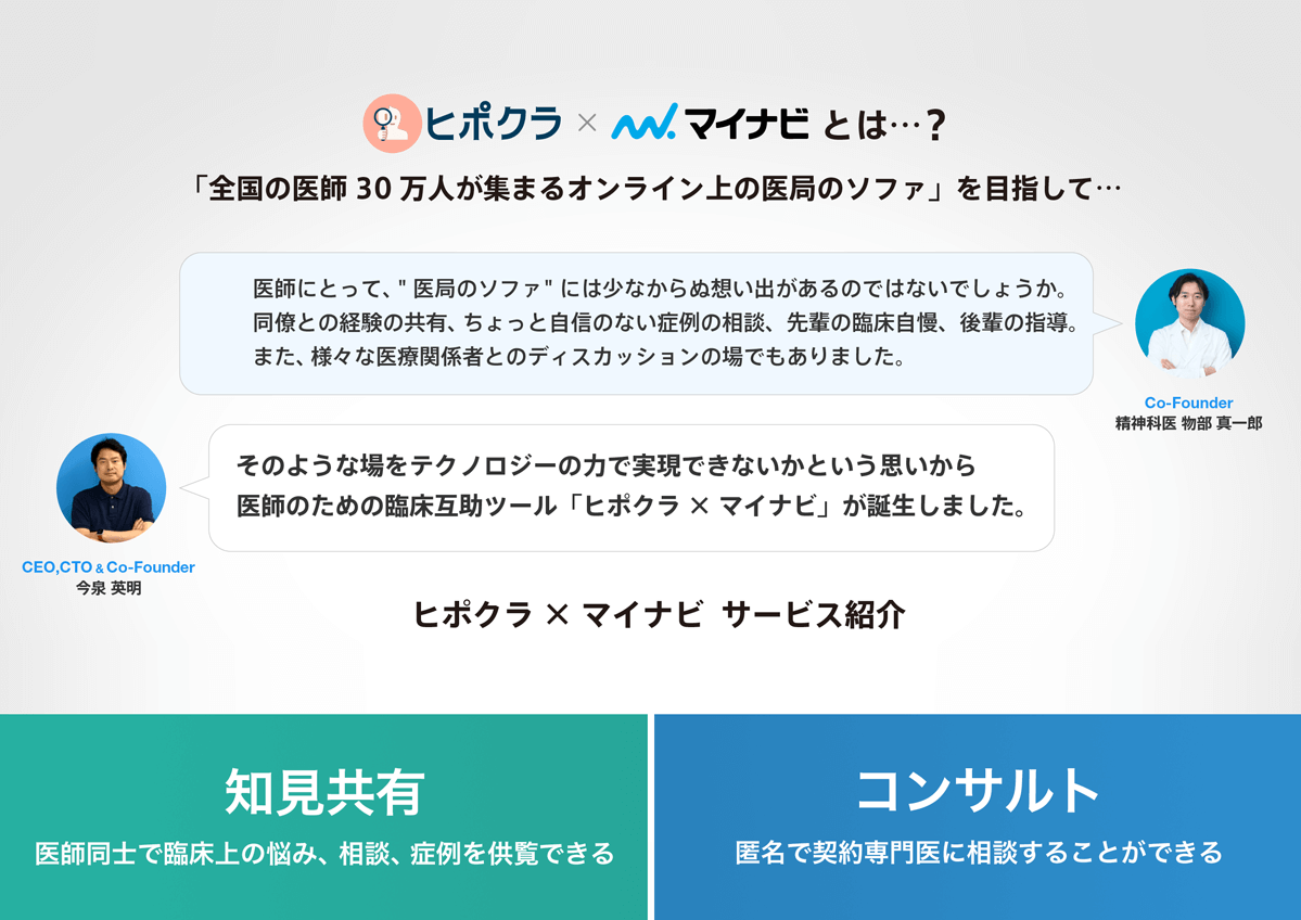
ヒポクラ x マイナビのご紹介

無料会員登録していただくと、さらに便利で効率的な検索が可能になります。