目的: 非外科的歯周治療 (NSPT) 後の唾液宿主細胞外小胞および非宿主細胞外小胞 (EV) の縦方向の変化を評価すること。 材料と方法: 刺激されていない唾液とプラーク (深いポケットから) を、ベースライン時と NSPT 後 3 か月および 6 か月目に、ステージ III/IV の歯周炎患者 25 名から収集しました。宿主唾液EVと非宿主EVは、それぞれ免疫親和性クロマトグラフィーとサイズ排除クロマトグラフィーを使用して単離されました。宿主 EV サイトカインはマルチプレックス フロー サイトメトリーによって評価され、非宿主 EV、唾液、プラークは 16S rRNA および真菌内部転写スペーサー (ITS) シーケンスによってプロファイリングされました。 結果: NSPT後3か月および6か月で歯周パラメータが大幅に減少し(ベースラインと比較)、宿主EVに関連するサイトカインであるインターロイキン-4(IL-4)、IL-12およびIL-9(唾液全体ではない)の増加が伴いました。両方の時点で、Filifactor、Parvomonas、Lentimicrobium 属、および Fusobacterium nucleatum、Filifactor alocis、Tannerella forsythia、Treponema denticola、Porphyromonas gingivalis、P. endodontalis、P. micra、および Lachnospiraceae 細菌種を含む細菌性 EV も減少しました。同様の減少は、トレポネーマ、タンネレラ、パルビモナス、T. レンギョウ、T. ソクランスキー、およびP. ミクロを含む歯垢および唾液分類群でも観察されました。6 ヵ月の時点で、マラセチア・アルナロケイの真菌EVは減少し、真菌カンジダ・パラプシロシスのEVは増加しました。 結論: NSPT は唾液の宿主および非宿主 EV 積荷の変化をもたらし、NSPT が歯周炎における疾患治療モニタリングのための非侵襲的ツールとして機能する可能性を示唆しています。
目的: 非外科的歯周治療 (NSPT) 後の唾液宿主細胞外小胞および非宿主細胞外小胞 (EV) の縦方向の変化を評価すること。 材料と方法: 刺激されていない唾液とプラーク (深いポケットから) を、ベースライン時と NSPT 後 3 か月および 6 か月目に、ステージ III/IV の歯周炎患者 25 名から収集しました。宿主唾液EVと非宿主EVは、それぞれ免疫親和性クロマトグラフィーとサイズ排除クロマトグラフィーを使用して単離されました。宿主 EV サイトカインはマルチプレックス フロー サイトメトリーによって評価され、非宿主 EV、唾液、プラークは 16S rRNA および真菌内部転写スペーサー (ITS) シーケンスによってプロファイリングされました。 結果: NSPT後3か月および6か月で歯周パラメータが大幅に減少し(ベースラインと比較)、宿主EVに関連するサイトカインであるインターロイキン-4(IL-4)、IL-12およびIL-9(唾液全体ではない)の増加が伴いました。両方の時点で、Filifactor、Parvomonas、Lentimicrobium 属、および Fusobacterium nucleatum、Filifactor alocis、Tannerella forsythia、Treponema denticola、Porphyromonas gingivalis、P. endodontalis、P. micra、および Lachnospiraceae 細菌種を含む細菌性 EV も減少しました。同様の減少は、トレポネーマ、タンネレラ、パルビモナス、T. レンギョウ、T. ソクランスキー、およびP. ミクロを含む歯垢および唾液分類群でも観察されました。6 ヵ月の時点で、マラセチア・アルナロケイの真菌EVは減少し、真菌カンジダ・パラプシロシスのEVは増加しました。 結論: NSPT は唾液の宿主および非宿主 EV 積荷の変化をもたらし、NSPT が歯周炎における疾患治療モニタリングのための非侵襲的ツールとして機能する可能性を示唆しています。
AIM: To evaluate the longitudinal changes in salivary host and non-host extracellular vesicles (EVs) following non-surgical periodontal therapy (NSPT). MATERIALS AND METHODS: Unstimulated saliva and plaque (from a deep pocket) were collected from 25 Stage III/IV periodontitis patients at baseline and at 3 and 6 months post NSPT. Host salivary EVs and non-host EVs were isolated using immunoaffinity, and size-exclusion chromatography, respectively. Host EV cytokines were assessed by multiplex flow cytometry, and non-host EVs, saliva and plaque were profiled by 16S rRNA and fungal internal transcribed spacer (ITS) sequencing. RESULTS: Periodontal parameters significantly decreased at 3 and 6 months post NSPT (vs. baseline), accompanied by increased host-EVs associated cytokines interleukin-4 (IL-4), IL-12 and IL-9 (not whole saliva). At both timepoints, bacterial EVs also declined, including the genera Filifactor, Parvimonas and Lentimicrobium and the species Fusobacterium nucleatum, Filifactor alocis, Tannerella forsythia, Treponema denticola, Porphyromonas gingivalis, P. endodontalis, P. micra and Lachnospiraceae bacterium; similar reductions were observed in plaque and saliva taxa, including Treponema, Tannerella, Parvimonas, T. forsythia, T. socranskii and P. micra. At 6 months, fungal EVs from Malassezia arunalokei were decreased and the fungal Candida parapsilosis EVs were increased. CONCLUSIONS: NSPT led to changes in salivary host and non-host EV cargoes, suggesting that they may serve as a non-invasive tool for disease treatment monitoring in periodontitis.
医師のための臨床サポートサービス
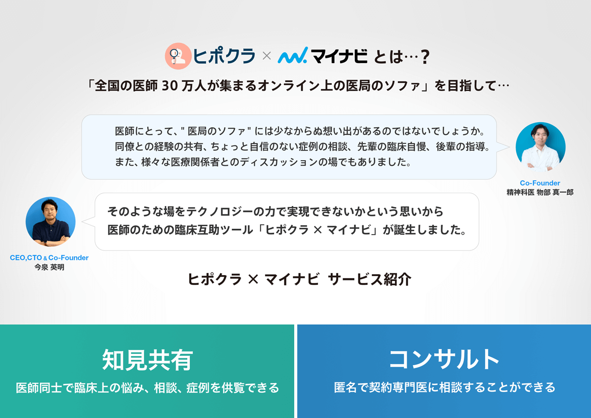
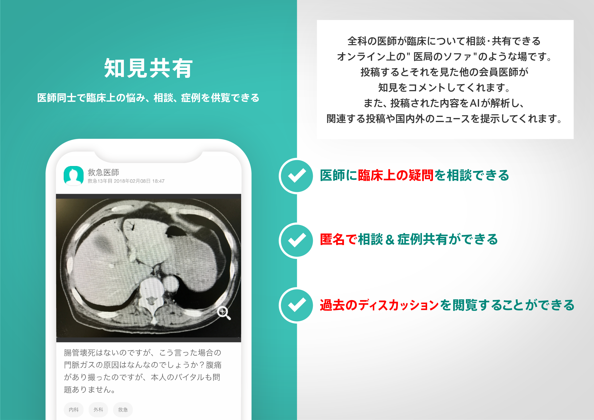
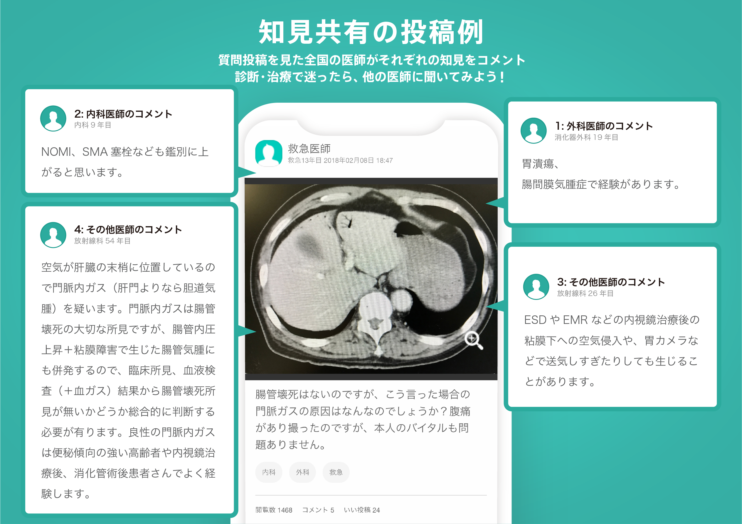
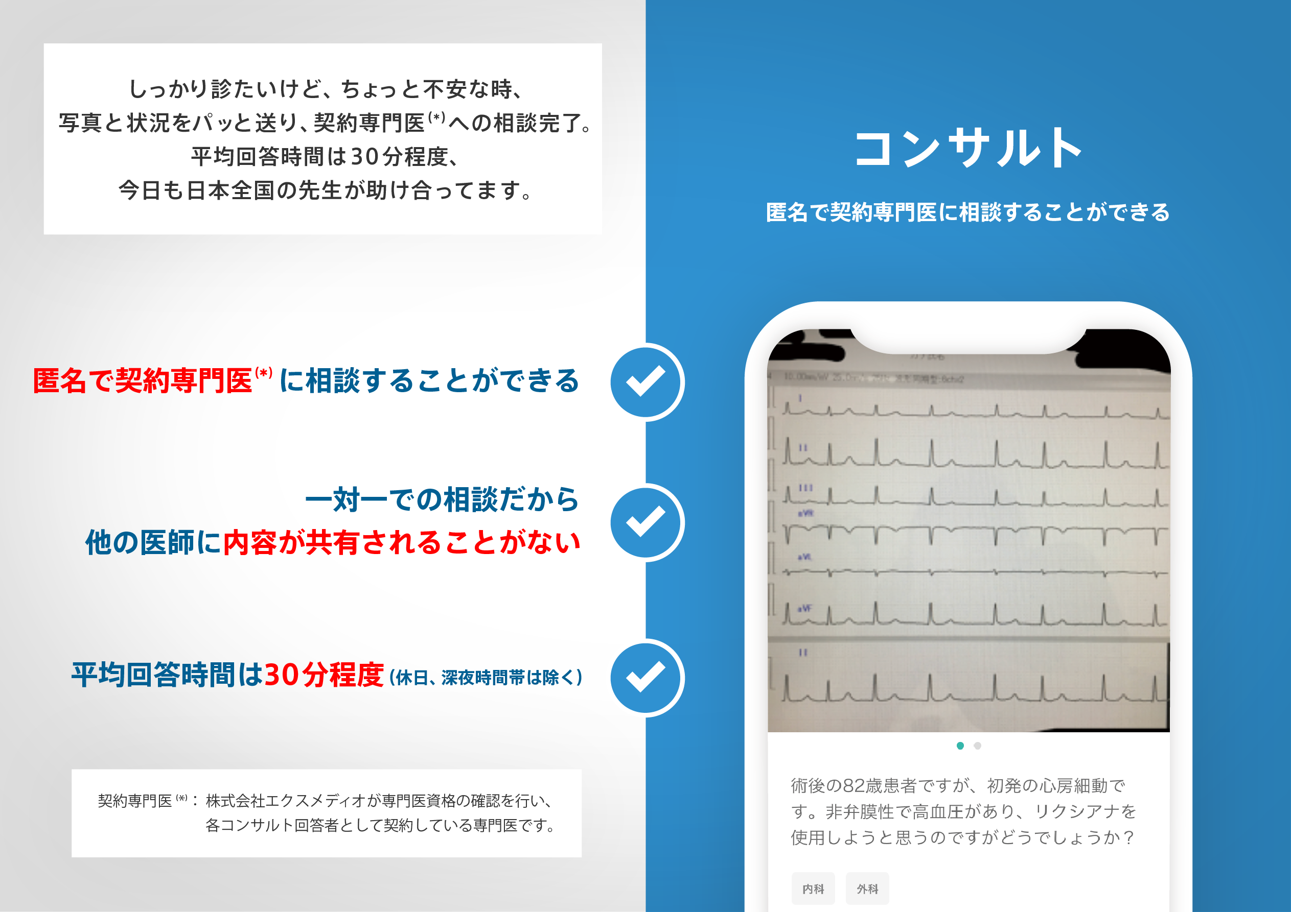
ヒポクラ x マイナビのご紹介

無料会員登録していただくと、さらに便利で効率的な検索が可能になります。