背景: 口腔悪性病変の早期発見は、その効果的な管理のために極めて重要です。病理組織検査は診断と治療計画のゴールドスタンダードですが、時間がかかり、専門家の評価と迅速な診断が不可欠なリソースが限られた環境では利用できないことがよくあります。人工知能 (AI) アプローチは、非侵襲的で迅速な代替手段を提供します。AI モデルは、早期発見を大幅に促進し、予後評価を改善し、個別化された治療戦略をサポートし、最終的に臨床転帰を向上させ、口腔腫瘍学における精密医療を進歩させることができます。 目的: この包括的なレビューは、口腔病変の悪性の可能性を検出および予測する際の AI モデルの有効性を評価することを目的としていました。 方法:「人工知能」、「口腔病変」、「系統的レビュー」というキーワードを使用して、コクランライブラリ、PubMed、Scopus、Web of Scienceのデータベースで包括的な検索を実施しました。特定された論文の適格性は、最初に、事前に定義された包含基準および除外基準に基づいて評価されました。その後、ジョアンナ・ブリッグス研究所(JBI)のチェックリストを使用して対象となる研究の方法論的な品質が評価され、品質基準を満たす研究がデータ抽出の対象に含まれました。データ抽出は 2 人のレビュー担当者によって独立して実行され、含まれたレビューの方法論的な品質は AMSTAR 2 ツールを使用して評価されました。 結果: AI と深層学習モデル、特に畳み込みニューラル ネットワーク (CNN) とハイブリッド アーキテクチャは、口腔扁平上皮癌 (OSCC) と口腔潜在悪性疾患 (OPMD) に対して優れた診断性能を実証しました。組織病理学ベースの AI モデルは、感度が最大 98%、特異度が最大 95%、診断オッズ比 (DOR) が 460.83 に達し、最高の診断性能を達成しました。臨床写真と光干渉断層撮影も、それぞれ約 91% と 88 ~ 90% の感度で高い診断精度を示しましたが、自己蛍光イメージングは比較的低いパフォーマンスを示しました。AI モデルは、経験の浅い臨床医を上回るパフォーマンスを示しながら、経験豊富な臨床医と同等の診断感度を達成しました。しかし、データセット、画像診断法、研究方法の不均一性により、研究間の直接比較は制限されていました。 結論: 含まれたレビューのほとんどにおける方法論的な制限にもかかわらず、AI モデル、特に組織病理学や臨床写真に適用された畳み込みニューラル ネットワークは、有望な初期結果と、制御された設定で専門家レベルの感度に近づく可能性を示しています。 トライアル登録: 論文は登録番号 CRD420251021811 で International Prospective Register of Systematic Reviews (PROSPERO) に登録されています。
背景: 口腔悪性病変の早期発見は、その効果的な管理のために極めて重要です。病理組織検査は診断と治療計画のゴールドスタンダードですが、時間がかかり、専門家の評価と迅速な診断が不可欠なリソースが限られた環境では利用できないことがよくあります。人工知能 (AI) アプローチは、非侵襲的で迅速な代替手段を提供します。AI モデルは、早期発見を大幅に促進し、予後評価を改善し、個別化された治療戦略をサポートし、最終的に臨床転帰を向上させ、口腔腫瘍学における精密医療を進歩させることができます。 目的: この包括的なレビューは、口腔病変の悪性の可能性を検出および予測する際の AI モデルの有効性を評価することを目的としていました。 方法:「人工知能」、「口腔病変」、「系統的レビュー」というキーワードを使用して、コクランライブラリ、PubMed、Scopus、Web of Scienceのデータベースで包括的な検索を実施しました。特定された論文の適格性は、最初に、事前に定義された包含基準および除外基準に基づいて評価されました。その後、ジョアンナ・ブリッグス研究所(JBI)のチェックリストを使用して対象となる研究の方法論的な品質が評価され、品質基準を満たす研究がデータ抽出の対象に含まれました。データ抽出は 2 人のレビュー担当者によって独立して実行され、含まれたレビューの方法論的な品質は AMSTAR 2 ツールを使用して評価されました。 結果: AI と深層学習モデル、特に畳み込みニューラル ネットワーク (CNN) とハイブリッド アーキテクチャは、口腔扁平上皮癌 (OSCC) と口腔潜在悪性疾患 (OPMD) に対して優れた診断性能を実証しました。組織病理学ベースの AI モデルは、感度が最大 98%、特異度が最大 95%、診断オッズ比 (DOR) が 460.83 に達し、最高の診断性能を達成しました。臨床写真と光干渉断層撮影も、それぞれ約 91% と 88 ~ 90% の感度で高い診断精度を示しましたが、自己蛍光イメージングは比較的低いパフォーマンスを示しました。AI モデルは、経験の浅い臨床医を上回るパフォーマンスを示しながら、経験豊富な臨床医と同等の診断感度を達成しました。しかし、データセット、画像診断法、研究方法の不均一性により、研究間の直接比較は制限されていました。 結論: 含まれたレビューのほとんどにおける方法論的な制限にもかかわらず、AI モデル、特に組織病理学や臨床写真に適用された畳み込みニューラル ネットワークは、有望な初期結果と、制御された設定で専門家レベルの感度に近づく可能性を示しています。 トライアル登録: 論文は登録番号 CRD420251021811 で International Prospective Register of Systematic Reviews (PROSPERO) に登録されています。
BACKGROUND: Early detection of malignant oral lesions is crucial for their effective management. While histopathological examination is the gold standard for diagnosis and treatment planning, it is time-consuming and often inaccessible in resource-limited settings where expert evaluation and rapid diagnosis are essential. Artificial intelligence (AI) approaches offer noninvasive and rapid alternatives. AI models can significantly facilitate early detection, improve prognostic assessment, and support personalized treatment strategies, ultimately enhancing clinical outcomes and advancing precision medicine in oral oncology. OBJECTIVE: The present umbrella review aimed to assess the effectiveness of AI models in detecting and predicting the malignant potential of oral lesions. METHOD: A comprehensive search was conducted in the Cochrane Library, PubMed, Scopus, and Web of Science databases using the keywords "artificial intelligence", "oral lesion", and "systematic review". The eligibility of identified articles was initially assessed based on predefined inclusion and exclusion criteria. Subsequently, the methodological quality of the eligible studies was appraised using the Joanna Briggs Institute (JBI) checklist, and studies meeting the quality standards were included for data extraction. Data extraction was performed independently by two reviewers, and the methodological quality of the included reviews was appraised using the AMSTAR 2 tool. RESULTS: AI and deep learning models, particularly convolutional neural networks (CNNs) and hybrid architectures, demonstrated excellent diagnostic performance for oral squamous cell carcinoma (OSCC) and oral potentially malignant disorders (OPMDs). Histopathology-based AI models achieved the highest diagnostic performance, with sensitivities of up to 98%, specificities of up to 95%, and diagnostic odds ratios (DORs) reaching 460.83. Clinical photography and optical coherence tomography also showed high diagnostic accuracy, with sensitivities of approximately 91% and 88-90%, respectively, whereas autofluorescence imaging demonstrated comparatively lower performance. AI models achieved diagnostic sensitivity comparable to experienced clinicians while outperforming less experienced practitioners. However, heterogeneity in datasets, imaging modalities, and study methodologies limited direct comparisons across studies. CONCLUSION: Despite methodological limitations in most included reviews, AI models, particularly convolutional neural networks applied to histopathology and clinical photography, show promising early results and potential to approach expert-level sensitivity in controlled settings. TRIAL REGISTRATION: Article Registered in the International Prospective Register of Systematic Reviews (PROSPERO) under registration number CRD420251021811.
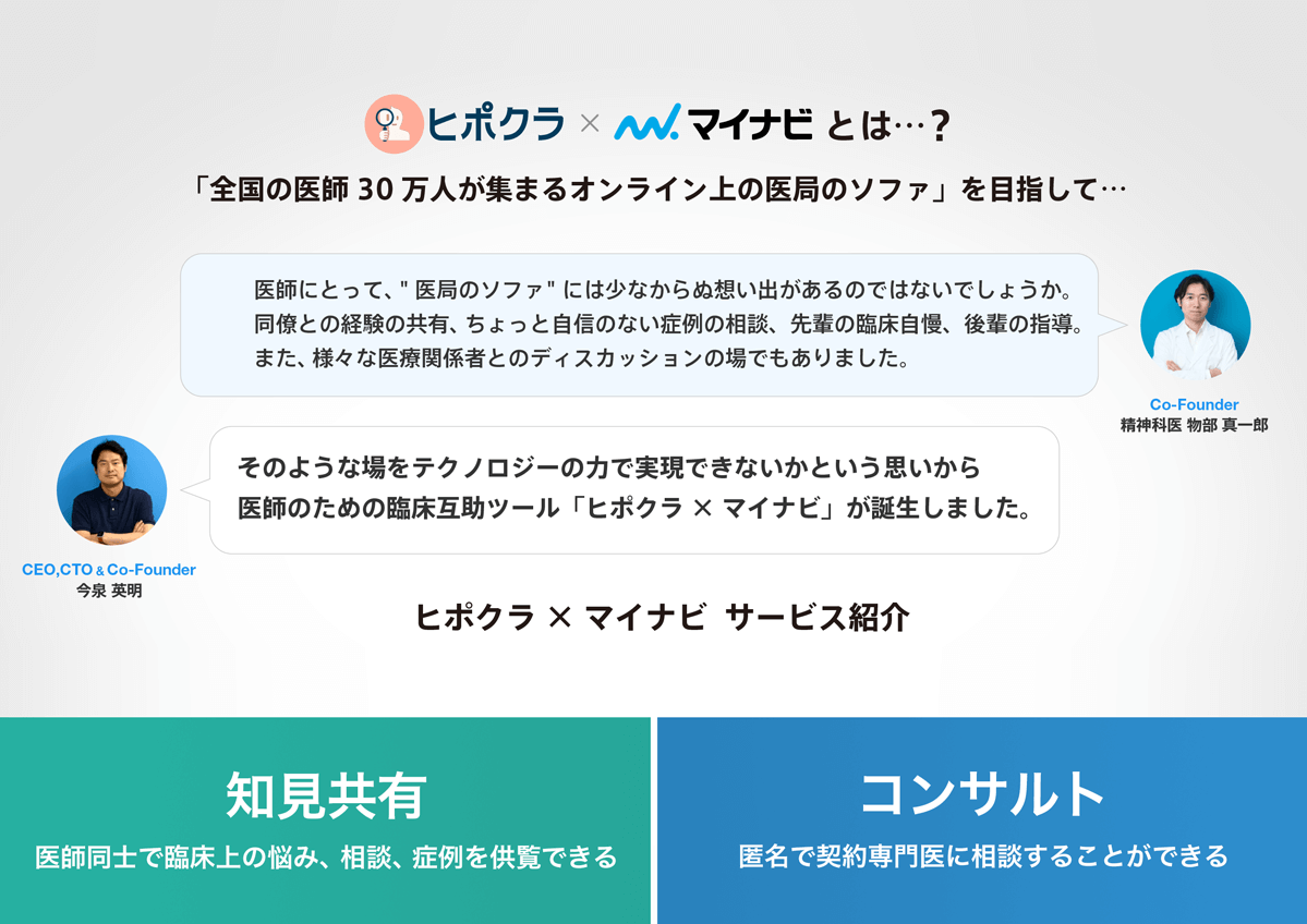
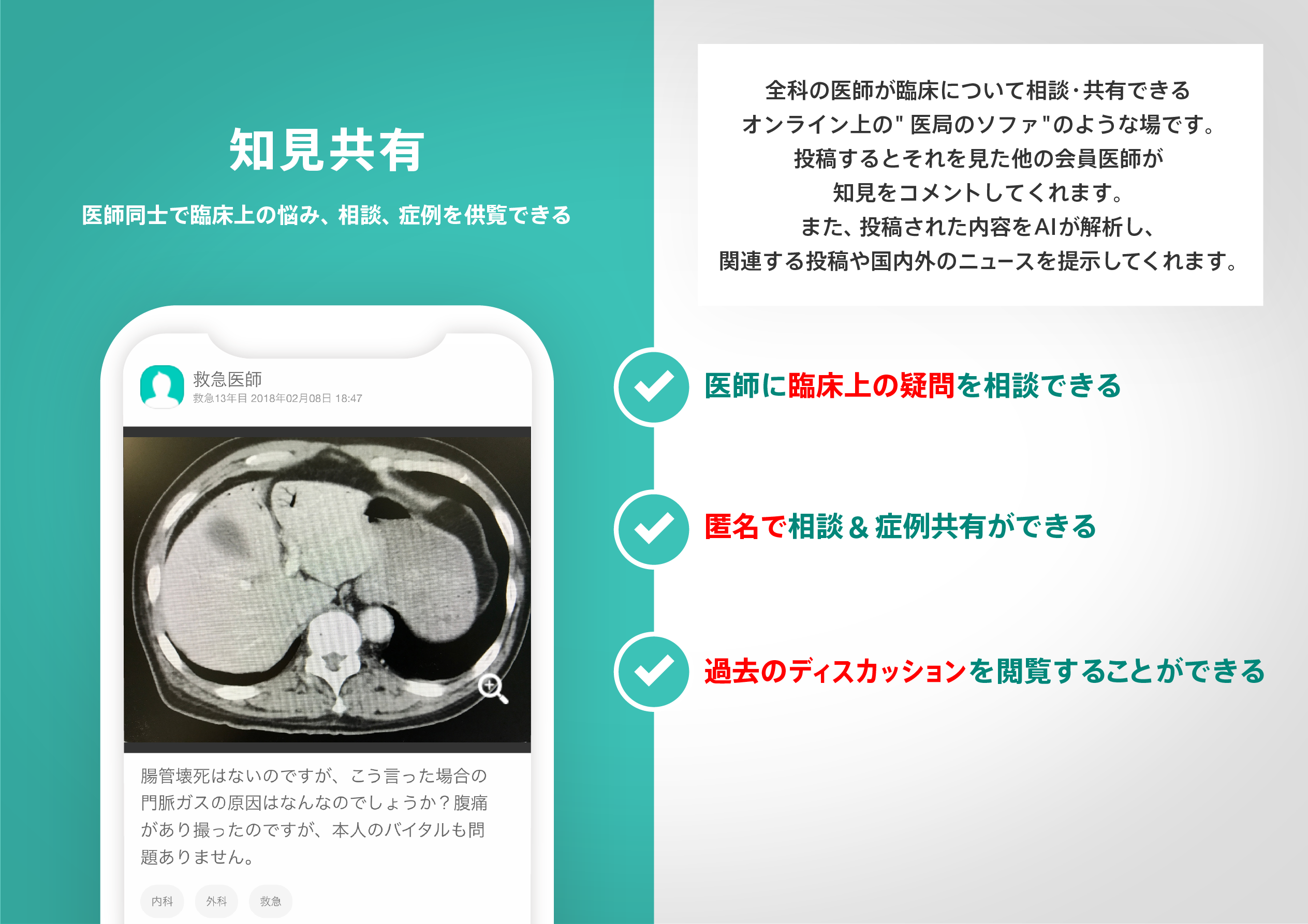
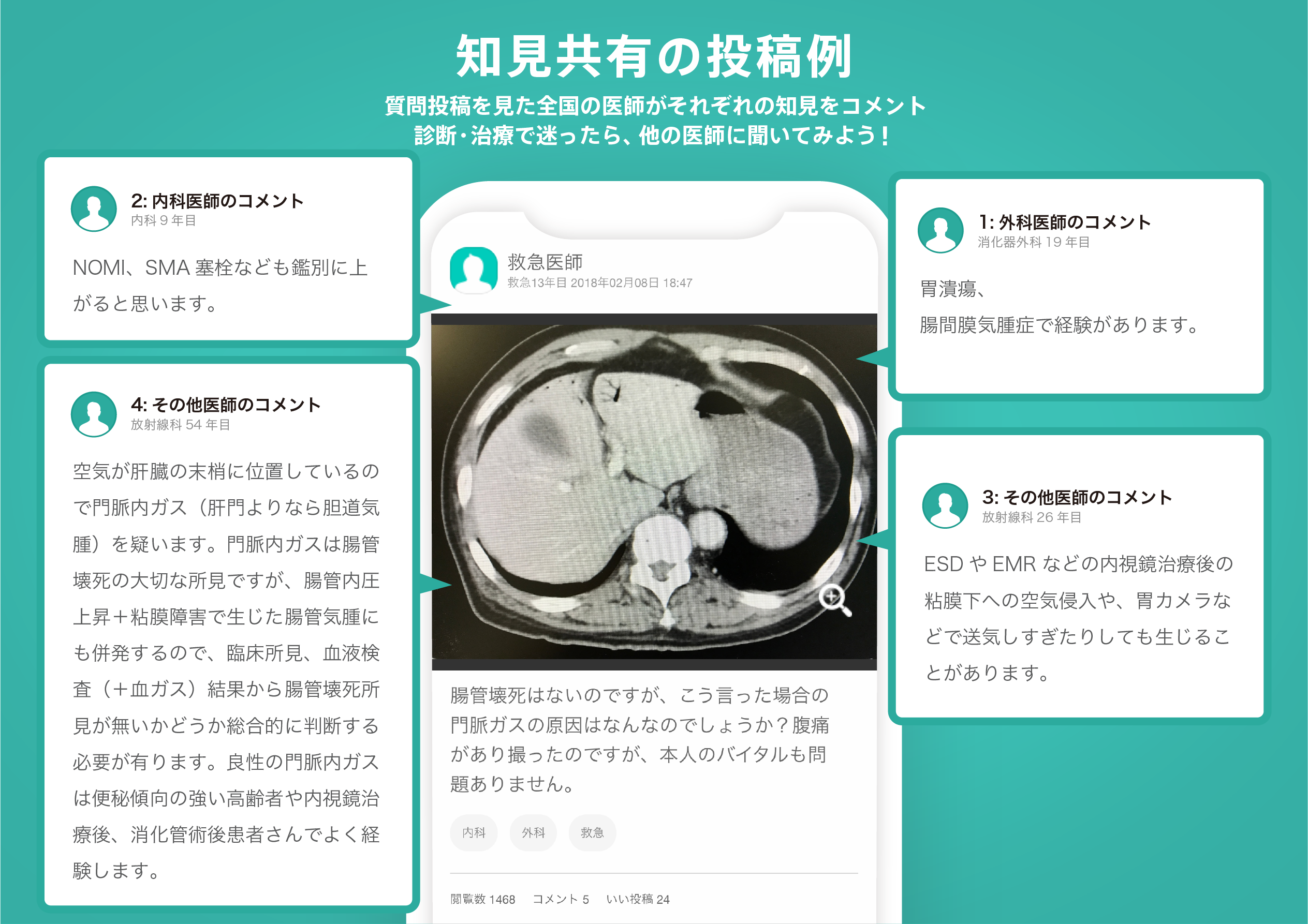
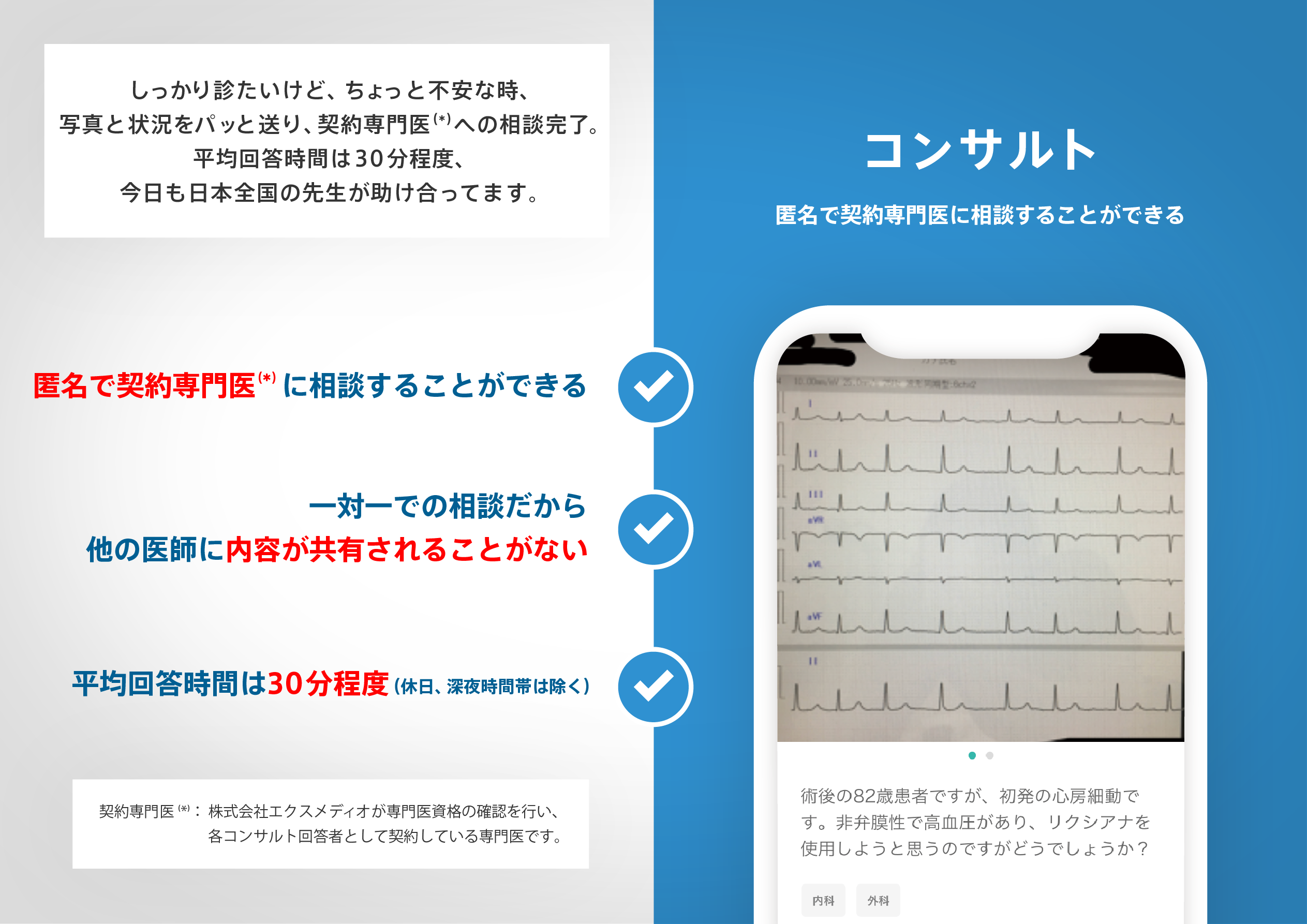
医師のための臨床サポートサービス
ヒポクラ x マイナビのご紹介

無料会員登録していただくと、さらに便利で効率的な検索が可能になります。